
Ultrasound is a non-invasive and widely used diagnostic tool. During the procedure, a healthcare professional uses a handheld device called a transducer to produce images on a screen.
Ultrasound scans may be used to:
- Confirm pregnancy
- Monitor fetal growth and development
- Determine the number of babies
- Evaluate the placenta and amniotic fluid
- Assess certain medical conditions
- Examine organs such as the thyroid and breasts
Types of ultrasound services include:
- Pregnancy Ultrasound
- 3D/4D Ultrasound
- NT/NB Scan
- Anomaly Scan
- Growth Scan
- Doppler Scan
- Fetal Echo
- TVS Scan
- Breast Ultrasound
- Thyroid Ultrasound
Pregnancy ultrasound helps healthcare providers:
- Confirm pregnancy
- Monitor fetal growth
- Assess the baby’s position
- Evaluate the placenta
- Monitor amniotic fluid levels
- Detect certain pregnancy-related concerns
Ultrasound scans do not use radiation. Instead, they use sound waves to create images of the developing baby.
Your healthcare provider will recommend ultrasound examinations based on your pregnancy and individual medical needs.
When Should I Have an Ultrasound?
Common pregnancy ultrasounds include:
- Early Pregnancy Scan
- NT/NB Scan
- Anomaly Scan
- Growth Scan
- Doppler Scan
- Fetal Echo
Unlike traditional ultrasound, which produces two-dimensional images, a 3D ultrasound combines multiple images to create a more detailed view.
A 3D ultrasound may help visualize:
- Facial features
- Hands and feet
- Body structure
- Certain developmental characteristics
Many parents choose a 3D ultrasound to create memorable keepsakes while also supporting prenatal care.
Benefits of 3D Ultrasound
- Enhanced visualization
- Detailed images
- Improved viewing experience
- Valuable addition to prenatal assessments
The timing and appropriateness of a 3D ultrasound should be discussed with your healthcare provider.
A 4D ultrasound builds upon 3D imaging by adding motion, creating a live video-like experience.
During a 4D ultrasound, you may observe:
- Facial expressions
- Hand movements
- Stretching
- Kicking
- Yawning
Many families choose 4D ultrasound as part of their pregnancy journey because it provides a unique opportunity to see their baby before birth.
Benefits of 4D Ultrasound
- Real-time imaging
- Enhanced bonding experience
- Detailed visualization
- Advanced diagnostic support
Your healthcare provider can recommend the appropriate timing for a 4D ultrasound based on your pregnancy.
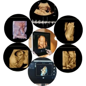
The quality of a 4D ultrasound image can depend on several factors, including:
- Gestational age
- Baby’s position
- Amount of amniotic fluid
- Maternal body type
During this stage of pregnancy, parents may be able to see:
- Facial features
- Hand and foot movements
- Smiles and yawns
- Stretching and kicking
However, every pregnancy is unique, and image quality can vary from person to person.
The NT scan is typically performed between 11 and 13 weeks and 6 days of pregnancy. During the scan, a healthcare professional measures the nuchal translucency (NT), which is the fluid-filled space at the back of the baby’s neck.
The NT scan may be used to:
- Assess the risk of certain chromosomal conditions
- Evaluate early fetal development
- Confirm gestational age
- Determine the number of babies
- Support first-trimester screening
An NT scan is usually combined with other assessments and should be interpreted by qualified healthcare professionals.
The NB scan is typically included as part of first-trimester screening between 11 and 13 weeks and 6 days of pregnancy.
Healthcare providers may use the NB scan to:
- Assess fetal development
- Support chromosomal screening
- Complement NT measurements
- Provide additional information during early pregnancy
NT and NB scans are commonly performed together to provide a more comprehensive assessment during the first trimester.
What Should I Expect During the Scan?
The procedure is similar to a standard pregnancy ultrasound and is generally painless. The duration of the examination may vary depending on the baby’s position and other factors.
An anomaly scan, also known as a mid-pregnancy or detailed scan, is commonly performed between 18 and 22 weeks of pregnancy.
During the scan, healthcare professionals may evaluate:
- The baby’s head and brain
- Face and spine
- Heart
- Abdomen
- Arms and legs
- Placenta
- Amniotic fluid levels
The purpose of the scan is to provide a detailed assessment of fetal development and identify any concerns that may require additional evaluation.
Why Is an Anomaly Scan Important?
An anomaly scan can help:
- Monitor fetal development
- Assess major body structures
- Support pregnancy management
- Provide reassurance to expectant parents
The timing of an anomaly scan is important because many of the baby’s organs and body structures have developed sufficiently to be evaluated.
An anomaly scan performed during this period may help assess:
- Brain development
- Heart structure
- Spine
- Limbs
- Placenta location
- Amniotic fluid
Your healthcare provider will recommend the most appropriate timing for the examination based on your pregnancy and medical history.
What Happens During the Scan?
During the procedure:
- Ultrasound gel is applied to the abdomen.
- Images of the baby are captured using a transducer.
- The examination typically takes 20–45 minutes, depending on the baby’s position and other factors.
A growth scan helps healthcare providers assess:
- The baby’s size and estimated weight
- Growth patterns
- Amniotic fluid levels
- Placental position
- Fetal movements and,
- Overall pregnancy progress
Growth scans are particularly useful in pregnancies that require additional monitoring or when there are concerns about fetal development.
Why Is a Growth Scan Important?
A growth scan can help:
- Monitor fetal growth over time
- Identify growth-related concerns
- Support pregnancy management
- Provide reassurance to expectant parents
Your healthcare provider will recommend the timing and frequency of growth scans based on your individual pregnancy.