
The Anomaly Scan: A Closer Look at What This Pregnancy Ultrasound Actually Checks
Somewhere between the 18th and 22nd week of pregnancy, most women are handed a referral slip for something called an “anomaly scan.” It’s a bigger appointment than the earlier ones longer, quieter, more technical. The sonographer spends far more time with the probe pressed against your abdomen, and there’s often a stretch of silence while measurements are taken that can feel unsettling if nobody has explained what’s actually happening on the screen.
This scan has a specific medical purpose, and understanding what it does and just as importantly, what it doesn’t makes the appointment far less anxiety-inducing.
Timing isn’t arbitrary. By this stage, the fetus has grown enough that its organs heart chambers, kidneys, cerebral ventricles, stomach, bladder, spine are large enough to be resolved clearly on ultrasound, yet there’s still enough amniotic fluid for the baby to move and reposition during the scan, giving the sonographer multiple viewing angles of each structure. Scan too early and structures are too small to assess reliably; scan much later and reduced fluid or fetal position can obscure views, particularly of the heart and spine.
This window also matters practically: if a significant structural anomaly is found, there is still time, depending on local laws and the family’s wishes, to have further tests, consult a specialist, or, in some cases, consider different options for the pregnancy while choices are still available.
What’s actually being measured ?
An anomaly scan isn’t a single “look” it follows a fairly rigid checklist, close to what’s outlined in ISUOG’s (International Society of Ultrasound in Obstetrics and Gynecology) practice guidelines for the mid-trimester scan. A typical protocol includes:
Biometry
Four standard measurements are taken to estimate gestational age and growth: biparietal diameter (BPD) and head circumference (HC) at the level of the thalami, abdominal circumference (AC) at the level of the stomach bubble and portal vein, and femur length (FL). These four numbers are plotted against growth charts.
Head and brain
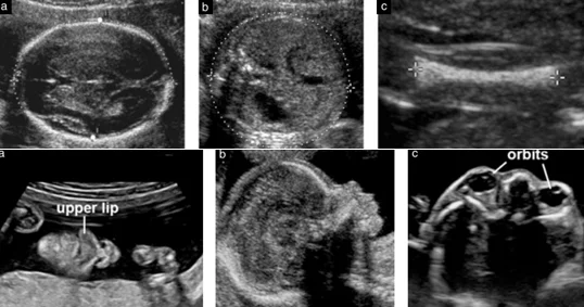
The shape of the skull, the cerebral ventricles, cerebellum, cisterna magna, and the midline structures (the falx and cavum septum pellucidum) are checked. This is where conditions like ventriculomegaly or structural brain malformations are picked up.
Face
A look at the profile, lips, and nasal bone; this is where cleft lip is usually identified.
Spine
Scanned in both longitudinal and transverse planes, checking the integrity of the skin covering over the spine, which is how open neural tube defects such as spina bifida are detected.
Heart
Arguably the most technically demanding part of the scan. The standard views include the four-chamber view, the left and right ventricular outflow tracts, and often the three-vessel view. Congenital heart disease is the most common category of major birth defect, and it’s also the one most frequently missed on ultrasound — detection rates for cardiac anomalies on a standard mid-trimester scan vary widely in published literature, often cited somewhere between 40–60% for isolated defects, which is why some pregnancies are referred on for a dedicated fetal echocardiogram if there’s a risk factor or a suspicious view.
Abdomen
Stomach, kidneys, bladder, and the insertion site of the umbilical cord into the abdominal wall are checked, along with confirmation that the diaphragm is intact (to rule out diaphragmatic hernia).
Limbs
Presence and length of the long bones in all four limbs, and a look at hands and feet.
Placenta and cord
Location of the placenta (importantly, whether it’s low-lying, which will need a follow-up scan later in pregnancy) and the number of vessels in the umbilical cord.
Amniotic fluid volume
Assessed either qualitatively or via a measured index.
The part that often gets misunderstood: “soft markers”
The appointment usually runs 30–45 minutes, sometimes longer if the baby is curled in a position that blocks key views cardiac views in particular are notoriously position-dependent, and it’s common to be asked to walk around, change position, or come back for a second look if a full assessment isn’t possible in one sitting. This isn’t a red flag; it’s routine. A moderately full bladder can help push the uterus into better view early on, though this varies by clinic. There’s no fasting requirement, no radiation involved (ultrasound uses sound waves), and nothing about the procedure itself carries risk to mother or baby.
If something is flagged
Most scans come back unremarkable. When a finding is flagged, the pathway typically depends on severity: minor or uncertain findings often just prompt a repeat scan in a couple of weeks to see if anything has changed or resolved; more significant findings are usually referred to a maternal-fetal medicine specialist for a detailed follow-up scan, sometimes alongside genetic counseling or invasive testing like amniocentesis if a chromosomal cause is suspected. The specifics matter enormously here a finding that sounds alarming in isolation (say, “echogenic bowel” or “single umbilical artery”) can carry very different implications depending on everything else seen on the scan, which is exactly why these results are interpreted by a clinician rather than read off a report in isolation.
A practical note for scheduling in Nepal
Anomaly scans here are typically performed by a radiologist or sonologist with obstetric ultrasound training, and it’s worth specifically confirming that the facility performs a detailed Level II / targeted scan rather than a routine dating or growth scan the equipment resolution and the sonographer’s training in fetal cardiac and neural views make a real difference to detection rates. If you have any risk factors a family history of congenital conditions, diabetes, a previous pregnancy with a structural anomaly, or an abnormal first-trimester screen it’s worth mentioning this before the scan so the sonographer can pay closer attention to the relevant structures.
This article is for general information and does not replace individual medical advice. If you have questions about your own anomaly scan results, please discuss them directly with your obstetrician or a maternal-fetal medicine specialist.