
How Breast Imaging Helps Detect Changes Early
“Breast scan” is a term patients use loosely for a handful of different imaging tests, and the confusion is understandable a mammogram and a breast ultrasound are both called scans, both look at the same organ, and both come up in the same conversations about screening and lumps, yet they work on completely different principles and are used for different reasons. Knowing which one you actually need, and why, makes the appointment far less confusing.
Mammography and ultrasound are not interchangeable
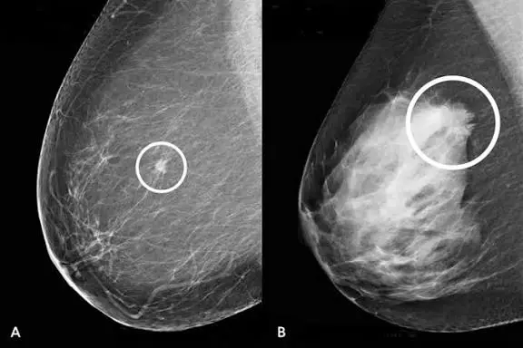
A mammogram is a low-dose X-ray of the breast, taken with the breast compressed between two plates to spread the tissue out for a clearer image. It is the only imaging test proven, at a population level, to reduce deaths from breast cancer through early detection, and it remains the backbone of routine screening in women without symptoms.
A breast ultrasound uses sound waves, not radiation, and is generally used to characterize a specific area for example, to work out whether a lump or an area flagged on a mammogram is solid or fluid-filled (a cyst), or as the primary screening tool in women whose breast tissue is too dense for a mammogram to read reliably. Ultrasound is also usually the first test used in women under 35, and often in pregnant or breastfeeding women, since it avoids radiation entirely.
In many cases the two are used together rather than as alternatives: a mammogram to survey the whole breast, and a targeted ultrasound to get a closer look at anything the mammogram picks up.
Why mammograms use compression ?
The flattening that makes a mammogram uncomfortable is not incidental to the test — it’s the mechanism that makes it work. Spreading the tissue reduces its thickness, which lowers the radiation dose needed, separates overlapping structures that could otherwise hide or mimic an abnormality, and holds the breast still to prevent motion blur. Compression typically lasts only a few seconds per image, and most women find it uncomfortable rather than painful; scheduling the scan for the week right after your period, when breast tissue is least tender, tends to make a real difference.
A standard screening mammogram takes two views of each breast one from above (craniocaudal) and one at an angle (mediolateral oblique) giving the radiologist two different perspectives on the same tissue, which is part of why a single view is never considered sufficient.
Breast density: the factor that changes everything
Breast tissue is a mix of fatty tissue, which appears dark on a mammogram, and fibroglandular tissue, which appears white unfortunately the same color a tumor appears. In women with dense breasts, a mammogram is effectively looking for a white spot against a white background, and its sensitivity drops meaningfully. This is not a rare situation: dense breast tissue is common, more so in younger women, and tends to decrease with age and after menopause.
Because of this, women who are told they have dense breasts are often advised to add a supplemental ultrasound (or in some cases an MRI) to their screening routine, since a normal mammogram in a dense breast carries somewhat less reassurance on its own than the same result in a less dense breast. If your report mentions breast density, it’s worth asking your doctor directly whether it changes your recommended screening approach.
Who should be screened, and how often
Screening recommendations vary somewhat between guideline bodies and are still an area of ongoing debate, but the broad shape is fairly consistent: average-risk women are generally advised to begin regular mammography somewhere in their 40s and continue on a schedule of every one to two years, continuing at least into their 70s. Women with a significantly elevated risk a strong family history, a known BRCA1/BRCA2 mutation, or a personal history of chest radiation at a young age are usually advised to start earlier, screen more frequently, and often add annual MRI to the routine.
It’s genuinely worth having this conversation individually with a doctor rather than following a generic age cutoff, since family history, prior breast biopsies, and personal risk factors can shift the recommended starting age and interval considerably.
What actually happens during a breast ultrasound
The technique is similar to any other ultrasound: gel is applied to the skin and a handheld probe is moved systematically across the breast and, often, the axilla (armpit), since lymph nodes there are checked as part of a complete breast evaluation. The whole exam usually takes 15–30 minutes, is painless, requires no preparation, and involves no radiation which is why it’s repeated as often as needed without the dose concerns that limit how frequently mammography is used.
If a lump is found, ultrasound is particularly good at answering one specific question quickly: is this a simple fluid-filled cyst, which is almost always benign and often needs no further action, or a solid mass, which usually needs a closer look, sometimes including a biopsy, before it can be characterized with confidence.
How results are reported: the BI-RADS system
Both mammograms and breast ultrasounds are reported using a standardized scale called BI-RADS (Breast Imaging Reporting and Data System), which categorizes findings from 0 to 6. In practical terms: BI-RADS 1 and 2 mean normal or clearly benign, with routine follow-up; BI-RADS 3 means probably benign, usually managed with a short-interval follow-up scan (often at 6 months) rather than immediate biopsy; BI-RADS 4 and 5 indicate suspicious to highly suggestive findings, where a biopsy is generally recommended; and BI-RADS 0 simply means the assessment is incomplete and additional imaging is needed before a category can be assigned. Seeing a number on your report rather than a plain-language verdict can be unsettling, but this system exists specifically to standardize how confidently a finding needs to be pursued, so it’s worth asking your doctor to translate the number rather than guessing at what it implies.
A finding doesn't mean cancer
This bears repeating because it’s the single most common source of unnecessary fear: the majority of findings flagged on a breast scan, including a fair number of BI-RADS 4 results, turn out to be benign on biopsy. Cysts, fibroadenomas, areas of fibrocystic change, and calcifications related to entirely benign processes are all common and are picked up regularly on routine screening. A recommendation for a follow-up scan or a biopsy is a step taken out of appropriate caution, not a sign that something serious has already been confirmed.
If you feel a lump between scheduled scans
Self-awareness of your own breasts noticing what’s normal for you remains useful even though formal monthly self-exam is no longer uniformly recommended as a stand-alone screening method. Any new, persistent lump, skin dimpling, nipple retraction or discharge (particularly if blood-tinged), or a change in breast size or shape should be brought to a doctor rather than waiting for the next scheduled screening, regardless of your age or how recently your last scan was normal. Most such findings turn out to be benign, but they warrant a targeted evaluation rather than reassurance from memory of an old scan.
Practical notes for your appointment
For a mammogram, avoid using deodorant, talcum powder, or lotion on the breasts and underarms on the day of the scan, since these can appear as artifacts on the image. Wearing a two-piece outfit makes undressing simpler, since only the upper body needs to be exposed. Bring copies or digital access to any prior breast imaging if you’re being scanned at a new facility radiologists rely heavily on comparison with previous scans to judge whether something has changed over time, and this comparison often matters more than any single scan read in isolation.
This article is for general information and does not replace individual medical advice. If you have questions about your own breast scan results or your personal screening schedule, please discuss them directly with your doctor.